About the Tool
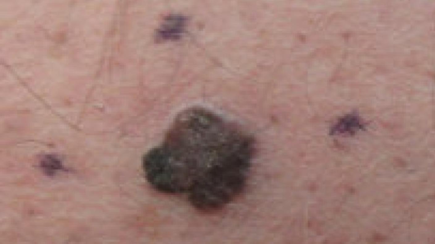
The Moles to Melanoma: Recognizing the ABCDE Features presents photographs in three main groups of pigmented lesions: common moles, dysplastic nevi (DN), and melanomas that arose from DN.
The DN section is subdivided into two broad categories: stable and fading, and evolving toward melanoma.
Each case series shows changes in an individual pigmented lesion over a number of years and across the spectrum of changes typically seen in U.S. melanoma-prone families.
We include a description of the “ABCDE” features for each type of pigmented lesion(moles, DN, and melanomas). Although the “ABCDE” rules were made for identifying early melanoma, they can also be used to describe DN.
About the Study
For more information about the study from which these cases were identified see the Clinical, Laboratory, and Epidemiologic Characterization of Individuals and Families at High Risk of Melanoma
Publications
Tucker MA, Fraser MC, Goldstein AM, et al. Melanomas and dysplastic nevi: A natural history atlas. Cancer. 2002.
Tucker MA, Halpern A, Holly EA, et al. Clinically recognized dysplastic nevi: a central risk factor for cutaneous melanoma. JAMA: The Journal of the American Medical Association 1997.
Acknowledgments
We thank the study participants for their many years of participation, their willingness to be photographed during skin examinations, and their generosity in allowing their pictures to be included in this resource.
We would also like to thank John Crawford and Mary King, NIH Clinical Center clinical photographers, for their expertise. The tool would not have been possible without the substantial commitment and cooperation of both the study participants and the clinical photographers.

