Computational and Physical Human Phantoms
REB investigators are developing models of human anatomy in both computational and physical formats, enabling them to estimate radiation doses to the human body and individual organs. Investigators use computational human phantoms for computing radiation dose by computer simulations, and then validate their results by using physical phantoms combined with radiation dosimeters.
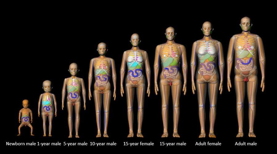
A "family picture" of computational phantoms, modeling a range of ages from newborn to adult.
Computational Phantoms
Two sets of computational human phantoms have been developed in collaboration with the University of Florida: a series of reference body size phantoms and a library of body size-dependent phantoms. These computational phantoms are combined with a Monte Carlo radiation transport technique to simulate a variety of radiation exposure scenarios and to estimate organ doses.
The computational phantoms are shared with the radiological community for research purposes as part of the NCIDose collection.
Physical Phantoms
The second version of human phantoms, called physical human phantoms, are made of stackable layers (called "slices"), each one dotted with a grid of holes into which tiny radiation sensors called dosimeters are inserted.
This kind of physical phantom can be used to provide accurate dose measurements when placed in a CT scanner or other source of radiation, but time and cost are substantial considerations.
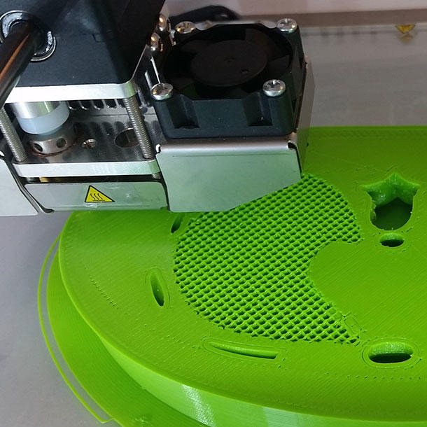
3D printer producing a "slice" of a phantom
In addition, physical phantoms available for purchase are limited in their sizes and features. So REB investigators are exploring the feasibility of producing 3D-print tissue blocks radiologically equivalent to human tissues. The 3D-printed tissue blocks could be used to customize physical phantoms to simulate features such as women's breast tissue and fat tissue for both male and females, enabling them to match computational human phantoms with varying body sizes.
Monte Carlo Simulation Methods
Monte Carlo is a class of numerical simulation methods used widely to solve complex physical and math problems. REB investigators are using three-dimensional Monte Carlo radiation transport. Detailed radiation propagation is modeled with Monte Carlo methods in computer simulations. This is a flexible and rigorous approach to simulate radiation transport within different materials, including human anatomy. The computational approach is extremely useful, particularly in dose reconstruction for large-scale epidemiological studies, where dose measurements are not feasible. REB investigators are using this method in dosimetry research to support several epidemiological studies of medical, occupational, and environmental exposure scenarios.
Uncertainties in Doses
Radiation doses calculated for epidemiological studies are associated with uncertainties because it is impossible to obtain precise and complete data for exposure assessment. For this reason, REB investigators are using Monte Carlo simulation methods to incorporate dosimetric uncertainties into radiation dose estimates. When incorporating uncertainty, some parameters associated with dose reconstruction are shared by cohort members and some are not. By using the two-dimensional Monte Carlo (2DMC) sampling method, the two types of parameters can be sampled separately and utilized for dose estimations. Multiple plausible doses (called realizations) are derived from the process, which then are used for risk analysis.
Radiation Measurements
REB investigators are using radiation measurements to validate radiation doses derived using computational methods. Instruments for radiation measurement available at REB are:
- Physical anthropomorphic phantoms representing a 5-year-old child and an adult
- Female breast phantoms specialized for dose measurement in breast radiotherapy
- Different types of dosimeters such as optically-stimulated luminescence dosimeter (OSLD) and ion chamber dosimeters; OSLD reading systems; and medical radiation spectrum measurement systems
Irradiation of physical phantoms equipped with dosimeters is conducted in collaboration with the NIH Clinical Center, the National Institute of Standards and Technology (NIST), and several medical facilities outside the NIH. Dose measurements using the equipment have been used to validate radiation doses computed for fluoroscopy, computed tomography, and radiation treatment procedures.
In addition, DCEG investigators are constructing physical anthropomorphic phantoms in-house using 3D printing techniques for more accurate and customized dose comparison with computational human phantoms.
For more information, contact Dr. Choonsik Lee or Dr. Vladimir Drozdovitch.