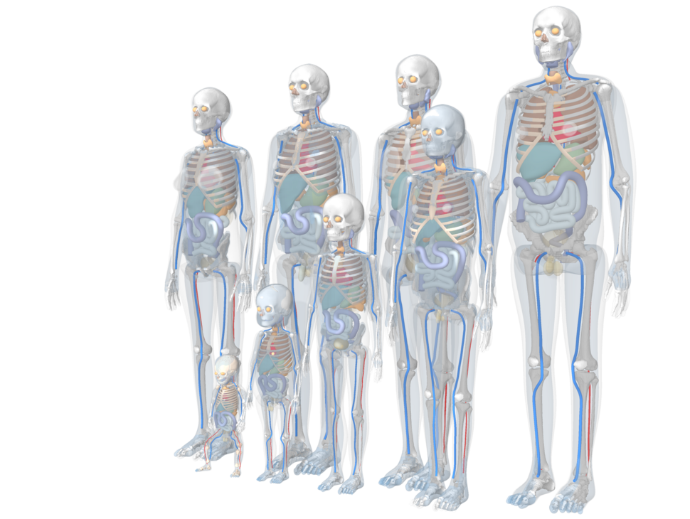
Family of reference-size computational human phantoms
PHANTOM is a computational human phantom library that provides a shared anatomical foundation for NCI Dose Tools and related radiation dosimetry research. Computational phantoms are three-dimensional representations of human anatomy used to estimate organ doses in medical radiation procedures, radiation protection, and epidemiologic studies. The models were developed from patient CT-based anatomical segmentation and modified to match reference anatomical and anthropometric data, producing realistic but standardized phantoms for organ-level dosimetry.
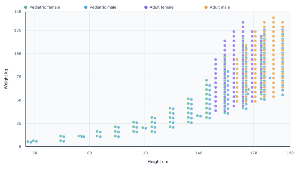
Distribution of height and weight of the size-dependent phantom library
The body size-dependent phantom library includes pediatric and adult models that represent both reference anatomy and a broad range of height and weight combinations. By selecting a phantom that more closely matches a patient or study population, NCI Dose Tools and related research applications can support more consistent organ dose assessment across different modalities and body habitus groups.
Resource Overview
- Provides anatomically detailed computational human phantoms for radiation dosimetry research.
- Supports pediatric and adult dose assessment using reference and size-dependent anatomy.
- Serves as a foundational data resource for NCICT, NCINM, NCIRF, and related dosimetry studies.
Additional information is available from related NCI Dose Tools GitHub sites:
- NCI Dose Tools GitHub Pages information site: more detailed tool information, worldwide use, and related publications.
- NCI Dose Tools GitHub technical repository: manuals, version history, technical documentation, and Q&A.
Access and Licensing
- Non-Commercial Research Use: There is no charge to use these resources for non-commercial research purposes. To request access, complete the Software Transfer Agreement form and submit the signed form to Dr. Choonsik Lee.
- Commercial Use: Commercial vendors and other commercial users should contact Dr. Kevin Chang of the NCI Technology Transfer Center to discuss trial access, licensing, and available integration options.